
Skull Model With Cervical Vertebra vs Plain Skull: BIX-A1013 Compared
Buy the BIX-A1013 if your course teaches the skull and the upper cervical spine together; buy a plain skull if you only need cranial landmarks. That is the whole decision. Physical models beat two-dimensional alternatives for anatomy learning, with a meta-analysis of comparative studies reporting a measurable advantage for physical models over other teaching media [1], so the question is not whether to buy a model but which level of detail your syllabus actually assesses.
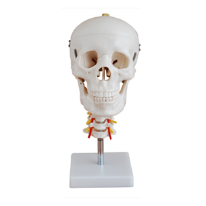
Quick facts
| Item | BIX-A1013 | Typical single-part skull |
|---|---|---|
| Composition | Skull plus C1–C7 cervical section | Skull only |
| Calvaria | Removable, cut for internal study | Usually fixed or 3-part |
| Neural detail | Hindbrain, spinal cord, cervical nerves shown | Cranial cavity only |
| Vascular detail | Vertebral artery, basilar artery, posterior artery | Not included |
| Mounting | Cervical section on a pedestal | Skull rests on a stand or flat base |
| Disassembly | Cervical vertebra can be separated from the cranium | Not applicable |
| Best for | Head-and-neck modules, neuroanatomy, palpation teaching | Introductory osteology, exam revision |
Where the extra cervical section earns its price
The skull does not sit in isolation in any clinical scenario a student will meet. Intubation, cervical immobilisation, whiplash assessment and headache referral all require the student to reason about the skull and the cervical spine as one unit. When the model shows the hindbrain, spinal cord and cervical nerves in continuity, a tutor can trace a pathway — from vertebral artery through the foramen magnum to the basilar and posterior arteries — without switching to a diagram.
That matters because the alternative is a mental jump. On a plain skull, the vertebral artery is a hole with a label. On the BIX-A1013, the artery, the cord and the nerve roots are physically present in the same object, which is the point at which students stop memorising and start explaining.
What the removable calvaria changes in a practical class
A removable vault converts a demonstration into a task. Students can open the cranium, identify the floor of the cranial fossae, then replace the vault and repeat the identification from the outside — the same model, two retrieval conditions. Lozano and colleagues made the case for physical prototyping in skull teaching on exactly this basis: tactile, three-dimensional objects support the spatial reasoning that flat images cannot supply [2].
The practical consequence for timetabling is small but real: a class of 20 needs roughly four to six models if students work in groups of four, and each rotation takes about 12 minutes.
Durability and handling notes
- PVC skull models tolerate repeated disassembly, but the cervical section is the wear point — check that the pedestal joint is metal-threaded, not glued.
- Store the calvaria separately if the class handles it frequently; chipped vault edges are the most common damage.
- Clean with mild detergent; alcohol wipes dull the painted arterial markings over time.
Procurement checklist
- Confirm the vertebral level. Ask whether the section runs C1–C7 or only C1–C3; the product data for BIX-A1013 states C1–C7.
- Check the arterial markings. Vertebral, basilar and posterior arteries should be painted, not moulded as unlabelled tubes.
- Ask for the pedestal specification. Weighted bases survive lab benches better than thin acrylic.
- Order spares for the calvaria cut. If the vault is removable, ask whether a replacement vault can be supplied separately.
- Plan storage. A skull on a pedestal needs shelf height, not drawer depth.
FAQ
Q1. Is this a full skeleton or a skull model?
A skull with a seven-vertebra cervical section on a pedestal. If you need a full skeleton, choose a separate product.
Q2. Can the calvaria be removed?
Yes. The cranial model is designed for disassembly so internal structures can be studied.
Q3. Which structures are shown beyond bone?
The hindbrain, spinal cord, cervical nerves, vertebral artery, basilar artery and posterior artery, according to the product specification.
Q4. Is it suitable for palpation teaching?
Yes for surface landmarks and cervical levels; it is not a soft-tissue simulator, so it cannot replace a task trainer for airway procedures.
Q5. What is the minimum order quantity?
Class sets are quoted per unit. Email chinonplus@adaanatomy.com with your cohort size and we will quote a class-set price with freight.
Q6. How does it compare with a 3-part skull?
A 3-part skull separates into vault and base for cranial study. The BIX-A1013 adds the cervical spine and vascular detail, which is what makes it useful for head-and-neck modules.
References
- Yammine K, Violato C. The effectiveness of physical models in teaching anatomy: a meta-analysis of comparative studies. Advances in Health Sciences Education. 2016. PMID 26459329.
- Lozano MTU, et al. 3D digitization and prototyping of the skull for practical use in the teaching of human anatomy. Journal of Medical Systems. 2017. PMID 28386686.
- McGaghie WC, et al. Does simulation-based medical education with deliberate practice yield better results than traditional clinical education? Academic Medicine. 2011. PMID 21512370.
Contact
For the specification sheet, class-set quotation and freight estimate: chinonplus@adaanatomy.com. Production follows ISO 45001 management practice; CE documentation is available for EU procurement.